

Joseph R. Anticaglia, MD
Medical Advisory Board
We take many things for granted when things are moving along smoothly. However, when things aren’t flowing normally, we become concerned. Why do I have a burning sensation on urination? Why is the color of the urine pink? Why do I have pain above my right hip and back? What’s causing my high blood pressure? My doctor told me I have a low red blood count.
The kidneys are vital, multi-task organs that contribute to our overall well-being. What follows is a look at the kidneys and how they function.
Kidneys
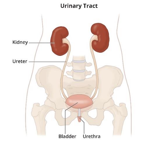
The kidneys are two bean-shaped organs located at the back of the upper abdomen, just below the rib cage. The ureters connect the kidneys to the bladder. The right kidney is slightly lower on the right side because of the position of the liver. They’re about the size of an adult’s fist, purplish brown in color due to the high vascularity of the kidneys. Each kidney weighs about one third of a pound (Fig.1).
The kidneys filter waste products and excess water from the blood to create urine. Waste and excess water are excreted from the body during urination. They remove waste products such as acids, muscle waste (creatinine) and nitrogen waste (urea).
They also reabsorb essential substances like glucose, amino acids, and water and return them to the bloodstream. The urinary system (tract) includes the kidneys and urinary pelvis. ureters, bladder and urethra (Fig1).
Anatomy Snapshot
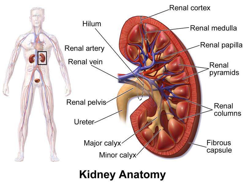
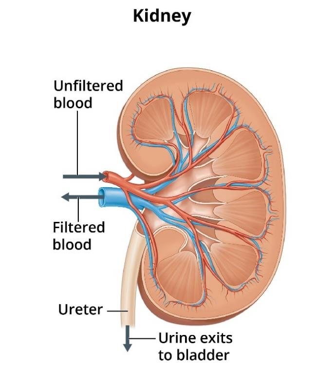
There’s an indentation in the middle of each kidney, called the renal hilum, which serves as the entry and exit point for the arteries, veins, nerves, ureter and lymphatics. Each kidney receives unfiltered blood from the renal artery. Filtered, cleansed blood is returned to the body when it leaves the kidney by way of the renal vein (Fig 2, 4).
On the outside of the kidney are three layers: 1) Dense connective tissue called renal fascia that holds the kidney in place. 2) Fatty, adipose tissue is the middle layer that protects the kidney from trauma—and 3) The third layer surrounding each kidney is called the fibrous or renal capsule.
On the inside (cross section) of each kidney are three main regions: renal cortex, medulla and renal pelvis (Fig2).
Renal Cortex and Nephrons
The renal cortex is located inside the fibrous capsule of the kidney. It is in this location where the vast majority of nephrons, cortical nephrons, are located and where blood filtration occurs. The nephrons are the functional units of the kidneys. Each human kidney has approximately one million nephrons (Fig.3). See Glossary — Nephrons
Medulla
The medulla is the inner part of the kidney. It contains the cone-shaped renal pyramids that help transport urine into a central cavity called the renal pelvis (fig 2). From the renal pelvis, urine flows to the tube-like ureters which transport the urine to the bladder. Finally, urine passes from the bladder through the urethra, another tube-like structure, to the outside during urination (Fig. 1).
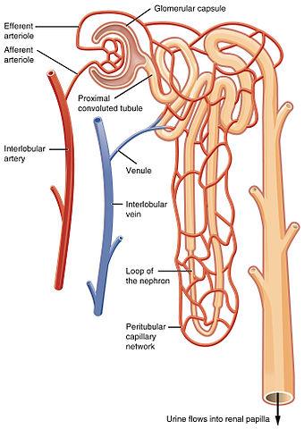
As unfiltered blood from the renal artery enters each kidney, it downsizes into a bunch of tiny vessels, the glomerulus. The glomerulus filters the blood and creates a filtrate (a liquid) in the space between it and Bowman’s capsule (Fig 3). As the filtrate travels through the renal tubules, urine is formed. And the two step process has been initiated; 1) excreting waste material and extra water into the urine, and 2) reabsorbing nutrients which are carried back to the body by the renal vein (Fig.4).
A future article will discuss how the kidneys are unsung powerhouses working to keep us alive.
References
- Ifeanyichukwu Ogobuiro; Faiz Tuma1. Physiology, Renal; StatPearls, July 25, 2022
- NIH Your kidneys and How They works
- J..L. Ssifter, and H. Chang; Disorders of Acid Base Balance: New Perspective; Kidney Disease (Basel), Jan2, 2017
- Edoardo Gronda et al; Glucose Metabolism in the Kidney; J. Am. Heart Ass. Nov.14, 2020
- Goss; Gray’s Anatomy; Lea and Febiger
- Medical Physiology Guyton Hall; Saunders
Glossary
Nitrogen waste — urea
Muscle waste — creatinine
Acid waste — hydrogen ions
Nephrons
Nephrons are the functional units of the kidneys. They work 24/7 to filter, clean the blood of waste products and excess water by creating urine, which is excreted from the body. They also reabsorb vital substances; nutrients needed by the body and return them to the bloodstream.
The main components of the nephron are the renal corpuscles, composed of the glomerulus and Bowman’s capsule. The glomerulus filters your blood and are found in innumerous numbers in the renal cortex.
The rest of the nephrons are called renal tubules which include the proximal tubule, loop of the nephron, and the distal tubule. Renal tubules remove liquid wastes via urination and return (reabsorb) needed substances to the bloodstream.
Cortical nephrons, with their short loops of the nephron, are located in the cortex of the kidney. When long loops of the nephron extend into the medulla, they are labelled juxtamedullary nephrons (Fig3).
This article is intended solely as a learning experience. Please consult your physician for diagnostic and treatment options.