

Joseph R. Anticaglia, MD
Medical Advisory Board
Glaucoma is a chronic eye disease that progresses slowly over time causing loss of eyesight. There is a buildup of pressure within the eye, because the eye’s drainage system is malfunctioning. A narrowing or complete blockage of the drainage system prevents fluid from flowing out of the eye in a normal fashion. This causes an excessive buildup of pressure within the eye that damages the optic nerve resulting in vision loss. It takes months to years before you notice eye problems, and the vision that has been lost cannot be restored.
Nancy started wearing eyeglasses when she was a teenager. Now in her early sixties, she has become accustomed to her yearly eye examination by her ophthalmologist, and was nonchalant when the doctor said: “I’m going to put a few numbing drops in both eyes, and I’ll be back in about 15 minutes to check the pressure.”
The doctor returned and moved the device called a tonometer into position to measure the intraocular pressure; that is, to measure the fluid pressure inside the eye. Nancy was startled when her doctor said, “I’m glad you keep your annual visits because today the intraocular pressure is elevated in both eyes with no damage to the optic nerve. This is an early stage of glaucoma.”
At this time, I suggest a few more tests, and ask you to return in three months to monitor the elevated eye pressure within the eye. I’ll call you about the results of the other tests. In the meantime, you need to take the eye drop medication that I’ll prescribe to be placed in each eye.”
And so, Nancy’s glaucoma journey began with many questions. Before she left the office she said “Doctor tell me more about glaucoma. What is glaucoma? What causes it? It sounds serious. Am I going to go blind?
The doctor answered Nancy’s questions by saying: “You’re not going to go blind. It is a serious condition and that’s why you must take the medication I prescribed, and keep the follow-up appointments. Now about glaucoma, and the doctor pointed to an eye chart on the wall.” (see figure below).
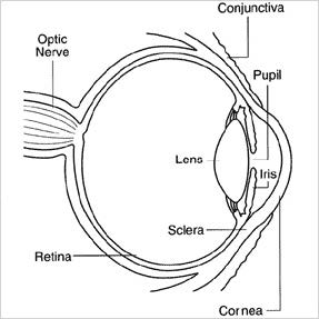
The doctor continued pointing to the chart and said: “First, I’d like to take a few seconds and talk about how we see. In general, light enters the eye through the pupil and travels through the lens on its way to the retina, which is located in the back of the eye.. The retina converts the light into electrical impulses which are sent to the brain via the optic nerve. The brain converts the electrical information into images.”
“Concerning glaucoma, it is an eye disease that causes irreversible blindness if not diagnosed and treated in a timely manner. It takes months to years before you notice eye symptoms. Many people have no clue that glaucoma is robbing them of their sight until they get the news after an eye examination. That’s why some people refer to this disease as the “Silent Thief of Sight.”
“Glaucoma happens when too much fluid builds up in the eye. The amount of fluid produced by the eye should equal the amount of fluid that is drained from the eye. The fluid buildup causes excessive pressure on the optic nerve. The pressure damages the nerve fibers of the optic nerve which now send inaccurate, faulty signals to the brain.”
Pointing to the chart, the doctor said, “The eye produces clear fluid in the back chamber of the eye, between the iris and the lens. The fluid then moves through the pupil into the front chamber between the iris and the cornea. Nancy, are you with me?
“No. Not really doctor… Just kidding. Just Kidding!” Both laughing…
“Anyway, if the drainage canal is partially or completely blocked, pressure builds up in the eye causing the lens to move backwards pushing the vitreous body backwards,” once again pointing to the chart, “damaging sections of the retinal nerves and optic nerve. The faulty signals which are sent to the brain causes loss of eyesight. People may notice loss of side (peripheral) vision initially, while retaining central vision also called tunnel vision.” (see image below.)
“Once the vision is lost, it’s gone. You cannot restore it. You need to take the eye drops and medication I prescribed as well as keep the follow-up appointments. Together, we’ll stop the progression of this disease, and prevent blindness. I’ve treated many patients in your situation, and they are doing well.
Two Major Types of Glaucoma
- Open angle (chronic) glaucoma
- Closed angle (acute) glaucoma


1. Open Angle (Chronic) Glaucoma
Open angle glaucoma is the most common type of glaucoma in the world. It accounts for ninety per cent of the cases in the United States. It’s a chronic disease that develops gradually, and may take many years before there is loss of vision. In virtually all cases of glaucoma, increased pressure within the eye damages the optic nerve’s ability to send needed electrical information to the brain for us to see.
The drainage angle in glaucoma refers to the location, the junction where the cornea and iris meet (see diagram above). In this type of eye disease, the entrance to the drainage canal is opened but the flow of fluid (aqueous humor) away from the eye slows down. There’s a partial blockage to the drainage system. Over time, a buildup of intraocular pressure damages the optic nerve and a person’s vision. The injured optic nerve fibers fail to transmit complete pictures to the brain.
Symptoms: Open Angle Glaucoma
This is a painless type of glaucoma that takes many years to develop. In the early stages of the disease, it may not be associated with loss of vision. As the disease progresses, people can experience bilateral peripheral vision loss (side vision loss) while retaining central vision, called tunnel vision (see above image). People might also complain of blurred vision, or rainbow-colored circles around bright lights.
Risk Factors
People with a family history of glaucoma, diabetes, and an elevated intraocular pressure are at a greater risk of developing glaucoma.
Diagnosis
Diagnosis of open angle glaucoma includes the identification of the following factors:
- Elevated intraocular pressure
- Visual field abnormalities (peripheral vision loss, tunnel vision)
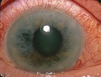
- Optic nerve cupping — The cup is a normal indentation of the optic nerve. Cupping indicates that there are large number of dead nerve fibers in the optic nerve. The cup becomes enlarged in patients with glaucoma.
Treatment
Eye drops and medications, laser and surgery have been used successfully to prevent vision loss. There is no cure for this type of vision loss. Early diagnosis and treatment are pivotal to prevent blindness.
2. Closed Angle (Acute) Glaucoma —Medical Emergency
Closed angle glaucoma is a medical emergency that requires immediate treatment. There is a sudden and complete blockage of aqueous humor at the drainage angle of the eye preventing fluid from flowing out of the eye. The blockage results in a quick, dangerous rise in eye pressure. It can happen when the pupil becomes dilated too quickly.
Infections, or trauma to the eye, for instance, can cause large amounts of substances like blood cells, and proteins to accumulate in the aqueous humor. The fluid with these substances totally block the drainage angle at its junction with the cornea and iris causing the sudden onset of symptoms.
Symptoms
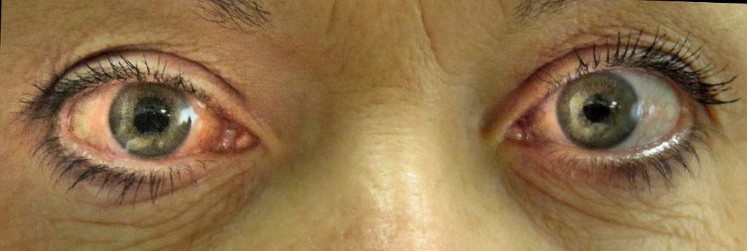
Acute closed angle glaucoma is characterized by sudden onset of severe eye pain, serious loss of vision, red eye, halos around bright lights, dilated pupil, nausea and vomiting. When timely treatment is initiated, patients with this type of glaucoma generally have favorable outcomes.
Other types of Glaucoma
Normal Tension Glaucoma
No one knows the exact reason the optic nerve becomes damaged when eye pressure is normal.
Congenital Glaucoma
Some infants are born with glaucoma or develop it within a few years due to abnormal drainage canals.
Secondary Glaucoma
This type of glaucoma can be caused by inflammation of the eye (uveitis) or eye injury.
Pigmentary Glaucoma
Happens when pigment granules from the iris accumulates in the eye’s drainage system
Glaucoma — after cataracts — it is the most common cause of vision loss in the U. S. Nine out of ten cases of glaucoma in this country is of the open angle type. Increase pressure within the eye damages the optic nerve, and if not treated in a timely manner causes irreversible blindness.
The only way to definitely diagnose glaucoma is by having a complete eye exam. Periodic examinations by your eye doctor every two years or less is the best way to prevent this type of vision loss. Acute closed angle glaucoma is an emergency that requires immediate management.
Glossary
Drainage Angle of the eye is the area where the cornea and the iris meet in the anterior chamber. The size of the angle determines how quickly aqueous humor fluid flows out of the eye. Several structures make up the drainage system at the angle of the eye, including the ciliary body, trabecular meshwork, and the canal of Schlemm.
Cupping, cup to disc ratio (C/D ratio) The optic nerve is divided into tenths, and the cup is compared to the entire optic nerve (optic disc) to obtain the cup-to-disc ratio. The normal cup to disc ratio is about 0.3 or about 1/3, which means the cup represents 1/3 of the optic discs height.
Closed angle glaucoma is also called angle closure glaucoma.
References
- Glaucoma; Wikipedia
- Kierstan Boyd; Reviewed By J Kevin McKinney, MD; What Is Glaucoma? Symptoms, Causes, Diagnosis, Treatment; Am Acad Ophthalmology, December 4, 2023
- John S. Cohen, MD and Harry A. Quigley, MD; Optic Nerve Cupping; Glaucoma Research Foundation, March 16, 2022.
- Babak Khazaeni et al; Acute Angle-Closure Glaucoma; StatPearls, November 26, 2023.
This article is intended solely as a learning experience. Please consult your physician for diagnostic and treatment options.