

Joseph R. Anticaglia, MD
Medical Advisory Board
A cardiac stress test evaluates the health of the heart. Sometimes called a treadmill stress test, it contrasts how the heart functions at rest compared to how it functions with activity. Either a treadmill or a stationary bicycle is commonly used by the person to put the heart under increased, physical stress.
An echocardiogram is a test that provides information about the action of the heart which is used to diagnose or monitor heart disease. It sends ultrasound waves into the heart that creates pictures which allow doctors to see the heart beating and pumping blood.
The pictures show the size and shape of the heart, if you have a blockage of the coronary arteries or problems with the heart valves.
The Echocardiogram Stress Test is a three part procedure. First, images of the heart are taken using the echocardiogram before the test begins.
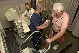
During the entire stress test, health care providers will monitor your performance. You will be attached to an electrocardiogram and your heart rhythm, breathing and blood pressure will be checked.
Second, when you’re on the treadmill, the level of cardiac stress is progressively increased by stepping-up the speed of the treadmill or increasing the steepness of the slope of the treadmill (its resistance) until you reach a target heart rate. The same idea applies to a stationary exercise bicycle.
The harder the body works during the stress test, the more energy the body needs to function well. The heart compensates by increasing the heart rate and the rate it pumps blood throughout the body. In order for the heart muscle to work efficiently, it needs to receive an adequate supply of blood from healthy arteries that go to the heart.
Narrowing and blockage of the coronary arteries can decrease the blood supply to the heart.
An irregular heartbeat can decrease the blood supply to the heart. These factors decrease the amount of oxygen available to the heart muscle and can lead to chest pain on exertion or precipitate a heart attack.
The third part of the procedure is to take post-exercise pictures immediately after completion of the treadmill. Valuable information is obtained by comparing images before and after exercising as well as reviewing the results of the electrocardiogram and blood pressure. Is there a blockage to one or more coronary arteries? Is the heart not getting enough oxygen? In other words, is there cardiac ischemia?
While at rest, a person with a blockage may have just enough blood circulating in the heart so as not to cause any symptoms. However, with physical activity, the heart muscle needs to work harder and might not get enough oxygen from reduced blood flowing around the blockage. In that case, exercise can cause chest pain and place you at a greater risk to suffer a heart attack.
If you’re not able to perform the treadmill test, another method to determine the blood flow to your heart is the nuclear stress test. It involves injecting a radioactive dye into your vein and taking images of your heart while you’re at rest and other images after the treadmill exercise is completed. The nuclear test helps diagnose coronary heart disease and may be useful as a guide in the treatment of this condition
Indications for a Stress Test
- To evaluate how your heart functions with exercise
- To diagnose if your symptoms of chest pain or difficulty breathing is heart related (coronary artery disease)
- To determine if you have an irregular heartbeat (cardiac arrhythmias)
- Initiate and evaluate treatment for patients with heart problems, for example, rheumatic heart disease, previous heart attack or congenital heart defect.
- Help provide guidelines for cardiac exercise programs
If the test is indicated, your doctor will give you special instruction concerning the treadmill stress test; for instance, what clothes to wear, use walking shoes, no smoking, when to stop eating and drinking before the test. Ask your physician about what medications are safe to take prior to the procedure and let him or her know if you’re using an inhaler.
You can stop the test at any time. If you have chest pain, feel dizzy, are fatigued or have severe shortness of breath inform the people monitoring you immediately. The personnel also have indications to stop the procedure such as an irregular heart rhythm or if the person’s blood pressure is too low or too high.
The total time for the test is approximately one hour and the actual exercise test time is ten to fifteen minutes. You’ll be monitored for about 15 minutes after you stopped exercising or until your heart rate returns to what it was before you started this test. Stress tests are generally safe, has proven to be valuable in diagnosing heart problems and helpful in guiding treatment.
References
- G Hillis; Basic transthoracic echocardiography; BMJ 2005
- Stephen G. Sawada et al; Prognostic value of a normal exercise echocardiogram; American Heart Journal; July 1990, Pages 49-55
- Thomas H Marwick; Stress echocardiography; Heart; January, 2003
- PeaceHealth; St. John Medical Center; Stress Testing/Stress Echocardiogram
- Gellish RL, Goslin BR, et al; Longitudinal modeling of the relationship between age and maximal heart rate. Med Sci Sports Exerc. May, 2007
This article is intended solely as a learning experience. Please consult your physician for diagnostic and treatment options.